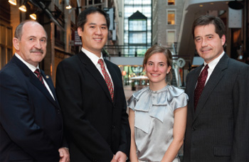
 De g. à d. : Le Dr Ron Smith, président de l'ADC; M. David Lee de l'Université de Toronto; Mme Jeanne Sansfaçon de l'Université Laval; M. David Hancin, vice-président et directeur général de Dentsply Canada.
Le Programme de recherche des cliniciens étudiants ADC/Dentsply 2010 a eu lieu à Montréal à l'occasion des Journées dentaires internationales du Québec (JDIQ) en mai. Tenu chaque année conjointement avec le Congrès annuel de l'ADC, ce concours national en recherche clinique invite des étudiants choisis dans les facultés de médecine dentaire agréées du Canada à présenter des démonstrations de leurs recherches cliniques devant des juges compétents. Le Dr Benoit Soucy, directeur des Affaires cliniques et scientifiques de l'ADC, et le Dr Louis Dubé, ancien président de l'ADC, ont agi comme juges cette année.
M. David Lee de l'Université de Toronto a été nommé lauréat du Programme 2010 pour son projet de recherche étudiant le lien entre les protéines morphogénétiques des os et les bisphosphonates. Son premier prix comprend un voyage tous frais payés au prochain Congrès annuel de l'Association dentaire américaine (ADA) à Orlando (Floride) où il présentera sa démonstration gagnante au cours du programme scientifique de l'ADA.
«Cette victoire est un reflet de tout l'appui que j'ai reçu», affirme M. Lee. «Mes superviseurs étaient toujours là pour m'aider et me guider et ma famille, surtout ma petite amie, m'a permis de conserver mon calme et de rester concentré à tout moment. Je ne saurais assez les remercier.»
Cette année marque un anniversaire spécial, étant la 40e année consécutive que Dentsply commandite le Programme de recherche des cliniciens étudiants. Ce programme réunit des étudiants de même mentalité de partout au pays et offre des occasions pour discuter et faire des découvertes. «La rencontre de confrères étudiants provenant de partout au Canada a été l'un des moments forts», ajoute M. Lee. «Le fait d'être en compagnie de gens aussi intelligents, compétents et sincères et de savoir que notre profession compte des êtres aussi formidables m'a rempli de fierté.»
La seconde lauréate à Montréal a été Mme Jeanne Sansfaçon de l'Université Laval. Son projet de recherche portait sur le lien entre les maladies parodontales et les infections intra-amniotiques. Mme Sansfaçon a intensément aimé l'expérience au complet et a ressenti un sens réel de camaraderie parmi les participants. «C'était intéressant de discuter ensemble nos projets de recherche ainsi que les divers problèmes que nous avons éprouvés», explique-t-elle. «Les questions posées par ces étudiants étaient très pertinentes et m'ont incité à réfléchir davantage à mon projet de recherche.» Pour avoir terminé au 2e rang, Mme Sansfaçon a reçu un prix de 1000 $ de Dentsply.
Prenant la parole lors de la distribution des prix, le Dr Ron Smith, président de l'ADC, a souligné que le concours vise à reconnaître les efforts des étudiants. «Au cours des 40 dernières années, ce programme a motivé des centaines de jeunes cerveaux brillants à étudier de nouvelles méthodes en vue de résoudre une multitude de problèmes de santé buccodentaire», a-t-il dit à l'auditoire assemblé. «Notre profession, ainsi que le public que nous servons, a appris et a profité des fruits de leur labeur.»
Chaque année, la Division canadienne de l'Académie Pierre Fauchard rend également hommage aux cliniciens étudiants qui participent au Programme ADC/Dentsply. L'Académie leur remet une bourse d'études en reconnaissance de leurs efforts spéciaux pour faire progresser l'éducation dentaire tout comme leurs carrières universitaires.
1re place
Le zolédronate en doses pharmacologiques inhibe l'effet ostéoinductif des BMP dans des cellules mésenchymateuses primaires humaines et des lignées cellulaires de souris
par David W.K. Lee, Université de Toronto
Conseillers : C. Clokie et S. Peel
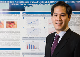 David Lee
Les protéines morphogénétiques osseuses (BMP) sont de plus en plus utilisées en clinique pour promouvoir la réparation osseuse. On remarque également qu'un nombre croissant de patients prennent des bisphosphonates pour le traitement de maladies osseuses (comme l'ostéoporose) ou de cancers avec métastases osseuses. Or, des concentrations micromolaires de bisphosphonates demeurent décelables dans les os (500 μM) et les tissus mous (1 μM) des semaines après l'administration de ces médicaments et on ignore les effets que ces taux de bisphosphonates peuvent avoir sur l'activité des BMP.
Les chercheurs ont mis en culture des cellules C2C12 de souris et des cellules mésenchymateuses primaires humaines (HUCPVC) en présence et en l'absence de rhBMP-2, avec des doses croissantes (0 à 100 μM) du bisphosphonate zolédronate (ZOL). La prolifération cellulaire a été évaluée à l'aide du colorant AlamarBlue. L'activité de la phosphatase alcaline (PhoA) et la teneur en protéines ont été mesurées pour évaluer la réponse des cellules aux BMP, et la toxicité du ZOL a été évaluée par la lactate-déshydrogénase (LDH). L'analyse de la variance à 2 critères de classification a été utilisée pour analyser les données.
Les données montrent que le ZOL a influencé l'ostéoinduction par les BMP en faisant intervenir 2 mécanismes distincts. À fortes doses (10 μM et plus), le ZOL a inhibé la prolifération cellulaire, réduit la teneur en protéines et élevé les taux de LDH dans les cellules HUCPVC et C2C12 (p 0,05). À faibles doses ( 1 μM) – auxquelles aucune différence significative dans la viabilité des cellules n'a été observée – le ZOL a inhibé l'augmentation de l'activité basale de la PhoA et de l'activité de la PhoA stimulée par les BMP (p 0,05). Les doses auxquelles ces 2 effets ont été observés se situent dans la gamme des concentrations de ZOL mesurées dans les os et les tissus mous des semaines après une seule injection, ce qui laisse croire que le traitement préalable par le ZOL pourrait influencer l'efficacité des BMP.
2e place
Association entre les maladies parodontales et les infections intra-amniotiques : état actuel des connaissances
par Jeanne Sansfaçon, Université Laval
Conseillère : F. Chandad
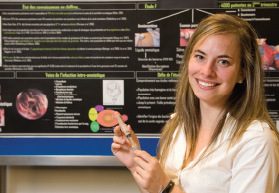 Jeanne Sansfaçon
Environ 1 enfant sur 10 naît prématurément. L'accouchement avant terme est la cause majeure de morbidité et de mortalité chez les nouveau-nés et présente un grand problème de santé publique.
Le foetus humain, pendant la majeure partie de sa gestation, baigne dans la cavité amniotique. Ce milieu est physiologiquement stérile. Or, il a été observé qu'une infection bactérienne de la cavité amniotique est présente chez près de 70% des femmes qui accouchent avant 30 semaines de gestation. L'origine de ces bactéries est diverse. Certaines pourraient avoir une origine ascendante ou descendante. Récemment, plusieurs évidences ont permis d'avancer l'hypothèse de l'origine buccale de ces infections intra-amniotiques. Cette hypothèse est émise car plusieurs espèces bactériennes isolées du liquide amniotique de femmes ayant accouché prématurément sont typiquement retrouvées dans la plaque dentaire. Il est connu que des bactéries présentes dans la cavité buccale peuvent envahir la circulation sanguine lors de micro-saignements, ce qui induit une bactériémie transitoire. Comme dans les cas d'endocardite bactérienne, ces bactéries buccales pourraient coloniser un autre milieu, en l'occurrence la cavité amniotique. Le défi est maintenant de faire la démonstration sans équivoque que les bactéries buccales retrouvées dans le liquide amniotique proviennent bel et bien de la cavité buccale de la patiente. L'objectif de cette présentation est de résumer l'état actuel des connaissances sur l'association entre les maladies parodontales et les infections intra-amniotiques et d'effectuer un survol de l'étude réalisée à ce sujet par la Faculté de médecine dentaire de l'Université Laval et le Centre Mère-Enfant du CHUL (CHUQ).
Les sommaires ci-dessous sont disponibles en anglais seulement.
Endodontic MTA materials support human mesenchymal cell attachment and differentiation
By Nelly Hashem, University of Western Ontario
Advisor: H. Perinpanayagam
 Nelly Hashem
Mineral trioxide aggregates (MTA) are widely used for pulp capping, perforation repair and root-end filling. However, little is known about the cellular response to MTA. The researchers’ objective was to determine if MTA supports human mesenchymal cell attachment, metabolic activity and osteogenic differentiation.
Human embryonic palatal mesenchymal (HEPM) cells were seeded onto grey (GMTA) and white (WMTA) MTA disks, as well as on titanium (Ti) and tissue culture plastic control surfaces. The number of cells that attached to each surface was counted with a hemocytometer and their metabolic activity was measured by MTT assay. Cell morphology and intracellular proteins were visualized by inverted immunofluoresecence microscopy. Gene expression was monitored by RT-PCR analysis.
Human mesenchymal cells attached and spread out on both grey and white MTA surfaces within 24 hours and proliferated over 72 hours of culture. Cell numbers and their metabolic activity were similar on the MTA and Ti surfaces after 24 hours of attachment, but increased more on the Ti after 72 hours of proliferation. The cells expressed Runx2 mRNA after 1, 3 and 7 days of growth on all of the surfaces. However, their expression of Runx2 protein was only detected after 3 days of differentiation on the MTA and Ti surfaces.
The researchers concluded that endodontic MTA materials support human mesenchymal cell attachment and metabolic activity, as seen on titanium implant surfaces. Furthermore, MTA supports Runx2 expression that is essential for osteogenic differentiation.
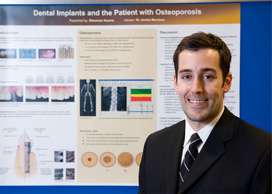
Dental implants and the patient with osteoporosis
By: Sheamus Kearns, Dalhousie University
Advisor: Dr. Archie Morrison
 Sheamus Kearns
Dental implants are becoming increasingly popular among clinicians and patients. In certain circumstances, they have become the treatment of choice for restoring edentulous spaces. Because osseointegration depends in part on the state of the host bone and its healing capacity, concerns have been raised about conditions affecting bone metabolism and its effect on implant prognosis. Success and survival rates for osseointegrated dental implants are well documented. However the survival and predictability of dental implants in patients with osteoporosis has been largely unknown.
The researchers performed a literature review to briefly explore the research pertaining to endosseous implant use in patients with osteoporosis and to provide some insight as to whether a diagnosis of this condition should contraindicate the use of these prosthetic devices in this particular patient population.
The researchers concluded that jaw bone quality, volume and density have an influence on the success of dental implants. However a diagnosis of osteoporosis based on bone density measurements outside of the jaws should not be reason to contraindicate the use of these prostheses.
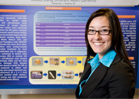
Microtensile bond strength of CAD/CAM blocks using different resin cements
By Jackie Samborski, University of Manitoba
Advisor: R. Roperto
 Jackie Samborski
Occlusal surfaces of extracted sound human molars were sectioned horizontally below the DEJ to produce dentin sections 3mm thick. Vita CEREC Blocks Mark-II (VT) and composite block Paradigm Z-100, 3M/ESPE (PZ) were sectioned into slices 3mm thick. Ceramic sections were surface-treated using a porcelain treatment kit. Each group was subdivided into 3 subgroups: the first was cemented to dentin using Panavia F, Kuraray (PA) as control; the second using self-adhesive resin cement SmartCem, Dentsply (SM); and the third using Calibra, Dentsply (CA). Bonded specimens were stored in water for 24 hours at 37°C then sectioned to obtain rods 1x1x6mm using a slow-speed diamond saw. The rods were then tested in a microtensile testing machine (Bisco). Data were calculated and statistically analyzed using ANOVA and Tukey's post hoc tests.
Means and SDs of μTBS (MPa) were: VT/PA: 11.66 (2.99); VT/SM: 5.08 (2.47); VT/CA: 13.84 (2.17); PZ/PA: 10.66 (5.52); PZ/SM: 5.85 (1.83); PZ/CA: 13.79 (3.11). ANOVA indicated significant differences among the cement groups and cement/block interaction (P<.01); and no significant difference among blocks only. Bond strength of PA groups (control) were consistently lower than CA groups but without statistical significance (P>.05) except when compared with SM groups, which was consistently lower (p<.01).
The researchers concluded that ceramic and composite CAD/CAM blocks had similar microtensile bond strength to dentin when 3 different resin cements were used.
Acknowledgements: VITA, 3M/ESPE and Dentsply.
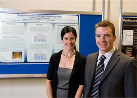
Synergistic inhibition of cariogenic bacteria by hops combined with fluoride
By Alison Schubert and Geoff McIntosh, University of Saskatchewan
Advisor: B. Ziola
 Alison Schubert and Geoff McIntosh
Numerous studies of Lactobacillus isolates have demonstrated that these bacteria are susceptible to the activity of hops. A recent study has shown that Streptococcus species are also sensitive to the effects of this antimicrobial. To date, no research has been conducted to determine whether any interactions exist between the antimicrobial activity of hops compounds and fluoride. The goal of the study was to determine whether a combination of fluoride and hops extract would work synergistically to inhibit caries-causing bacteria.
Experimental bacterial isolates, including several isolates of Streptococcus mutans, Streptococcus sanguinis, Streptococcus salivarius, Lactobacillus acidophilus and Lactobacillus casei, were grown in MRS media until visibly turbid. Gradient plates containing agar only (control), agar and hops, agar and fluoride and agar, hops and fluoride, were inoculated using the edge of a microscope slide along the gradient of each plate. The plates were incubated in a candle jar at 30°C for 48 hours, after which the distance of bacterial growth up each gradient was compared.
The researchers determined that not only does a combination of fluoride and hops compounds appear to elicit greater antimicrobial activity against hops-susceptible isolates, it also shows activity against isolates resistant to the activity of hops. Further research in the form of more widespread screening of oral, caries-causing bacteria is needed. These early results are encouraging for the inclusion of hops and fluoride combinations into antimicrobial dental products.
Rehabilitation by removable prosthesis of a deep class II occlusion
By Tudor-Ioan Stiharu, University of Montreal
Advisor: G. Gauthier
 Tudor-Ioan Stiharu
Fifteen percent of the population lives with a skeletal class II occlusion. At an advanced age, some of these people require fixed or removable prostheses in order to properly function.
A patient presented with an immediate full upper and defective Kennedy class 1 lower partial dentures to be rehabilitated with a permanent full upper and functional lower dentures. The patient exhibited a skeletal class II occlusion with an under bite of more than 10mm.
After diagnosis, initial imprints were taken and individual imprint trays were crafted. The cast metal frame of the lower partial denture was judged adequate and was reused since it had been made less than a year prior to presentation. Final polysulfur imprint of the upper maxilla and Korrecta wax tertiary imprint of the edentulous posterior mandible were acquired. Final models were obtained and occlusion rims were adjusted while vertical dimension was established. Centric relation and face bow were recorded for intermaxillary relations. Models were mounted on a Hannau Wide View articulator and prosthetic acrylic teeth were placed on occlusion rims. A double row of posterior teeth was used for the upper maxilla. Aesthetic trial was performed and prostheses were cooked. Final occlusal adjustments were done and patient received prostheses.
The researchers achieved clinical success, with the patient being very satisfied functionally and aesthetically. They believe this method should be considered when attempting to restore deep class II edentulous or partially edentulous patients with removable prostheses.
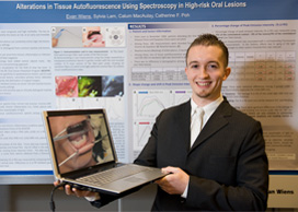
Alterations in tissue autofluorescence using spectroscopy in high-risk oral lesions
By Evan Wiens, University of British Columbia
Advisor: R. Shah
 Evan Wiens
High-risk oral lesions (HRL) are sometimes difficult to discriminate from reactive lesions (RL) commonly seen in community settings. Alteration in tissue autofluorescence is one promising approach for the early detection of HRLs. The objectives of this study were to collect autofluorescence spectra from normal mucosa, HRLs and RLs under different light excitation wavelengths and to compare the changes in tissue autofluorescence among these lesions.
Patients were recruited from the dysplasia clinics of the BC Oral Cancer Prevention Program. Spectroscopic measurements were taken using a probe with 3 excitation wavelengths (blue [436nm], violet [405nm] and UV [350-360nm]). The loss of Peak Emission Intensity (%ΔPEI) was measured by comparing the PEI of the lesion and contralateral normal areas. Differences in the %PEI between groups were compared.
From June to November 2009, 186 spectroscopic measurements were recorded from 31 patients. When comparing the %ΔPEI between cancer and dysplasia, there was an increased loss in the cancer group at all 3 excitation wavelengths. Almost the same loss was observed between cancer and RL. Importantly, there was a significant difference in %ΔPEI between dysplasia and RL under 436nm and UV excitations, but no difference under 405nm excitation.
This is the first study to use 3 different excitation wavelengths to examine oral cancerous, dysplastic and reactive lesions. This technology has shown potential to provide an objective and sensitive approach to distinguish between precancer and reactive lesions.
Toutes les photos sont une gracieuseté de tecklesphoto.com