A 32-year-old man was referred to the oral diagnosis and radiology department of a university dental faculty for investigation of gingival bleeding. His medical history was unremarkable, and he had not been taking any medications. Intraoral examination revealed marked gingival hyperplasia (Fig. 1a). The results of a complete blood count indicated no significant abnormalities, with only slight decreases in red blood cells, hemoglobin and hematocrit and a slight elevation of lymphocytes. For further analysis and confirmation of these results, another complete blood count was performed 2 days later, at which time levels of free thyroxine (T4) and thyroid-stimulating hormone (TSH) were also determined. The patient's T4 level was low, whereas his TSH was elevated. Levels of antibodies to thyroid peroxidase (titre 1049) and thyroglobulin (titre 1075) were highly elevated. Ultrasonography revealed that both lobes of the thyroid gland were smaller than normal, with lobulated contours. The echotexture was pseudonodular. Thickening of the hair and excessive hairiness of the face were detected during an extraoral examination, and the patient stated that he had gained weight (Fig. 1b). Desquamation of the patient's back was evident (Fig. 1c). Incisional biopsy of the gingiva, followed by histopathologic examination, was performed.
 Figure 1a: Photograph of a 32-year-old male patient at the time of presentation shows remarkable gingival hyperplasia. The gingival tissue between teeth 11 and 12 resembled an epulis.
 Figure 1b: Extraoral examination showed excessive hairiness of the face.
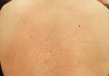 Figure 1c: Desquamation was observed on the patient's back.
What is the diagnosis?
Differential Diagnosis
Gingival hyperplasia has been associated with a variety of factors, including medications, inflammation and systemic disorders.
Over the past few years, the list of medications having a documented association with this type of gingival overgrowth has increased. This list includes anticonvulsant drugs, potent immunosuppressive agents and certain antihypertensive drugs. The fact that phenytoin, cyclosporin and calcium-channel blockers are all calcium antagonists suggests a common pathogenetic basis for gingival hyperplasia influenced by these drugs.1
Gingival hyperplasia may also occur when microorganisms present in bacterial plaque on tooth surfaces invade the gingival sulcus. The affected gingival tissues are edematous, their consistency is soft, and they may bleed upon gentle probing.2,3
Several systemic conditions may lead to gingival hyperplasia. For example, in patients with acute monocytic, lymphocytic or myelocytic leukemia, the gingival tissues may be enlarged, edematous, soft and tender to the touch, and they tend to bleed easily.
Inflammation of dental plaque leading to gingival hyperplasia may occur in association with some hormonal conditions (e.g., pregnancy, puberty or hypothyroidism), nutritional conditions (e.g., vitamin C deficiency) or nonspecific conditioned hyperplasia (e.g., pyogenic granuloma).2-6
Recent periodontal studies have suggested a mild to moderate association between human periodontal disease and some systemic disorders such as Hashimoto disease.3,5 Hashimoto thyroiditis is the most common cause of primary hypothyroidism. Medical conditions associated with hypothyroidism include hypercholesterolemia, hyponatremia and anemia. Hashimoto thyroiditis may be associated with other autoimmune diseases such as pernicious anemia.7
Among previously reported oral findings of hypothyroidism, there has been only one case report of gingival hyperplasia.8 In this situation, the clinical consequences of altered gingival microcirculation could compromise the first line of defence against periodontal disease. Contact of lymphocytes and plasmocytes with the vascular wall confirms that vascular factors trigger damage to the periodontium.6 Microvascular alterations in the interdental papilla may represent a deficiency in local periodontal defence mechanisms and may consequently be associated with periodontal disease.3
Definitive Diagnosis and Treatment
Gingival fibromatosis was diagnosed in this patient on the basis of his medical history and clinical features. In addition, the ultrasonographic findings and levels of antibodies to thyroid peroxidase and thyroglobulin led to a diagnosis of Hashimoto disease. More specifically, the patient had not been using any medications that would have induced the gingival enlargement. In addition, the gingival enlargement was localized, the patient had gained weight, and increased hairiness of the face was present, along with thickening of individual hairs. Desquamation of the skin was another diagnostic clinical feature. The complete blood count indicated anemia, and the T4 and TSH levels were abnormal. Pseudonodular echotexture of the thyroid glands on ultrasonography, as observed in this patient, is characteristic of Hashimoto disease, as is elevation of antibodies to thyroid peroxidase and thyroglobulin.
Treatment with levothyroxine sodium (0.1 mg per day) for hypothyroidism and vitamin B12 (1000 µg per month) for anemia was initiated. The patient was continuing to receive these therapies at the time of writing, in late 2011. No periodontal treatment was applied. By 2 months after diagnosis, the patient's thyroid status had returned to normal, and gingival remission was observed (Fig. 2). During a follow-up examination 1 year later, total remission of the gingival hyperplasia was observed (Fig. 3). At that time, periodontal therapy consisted of scaling only. After 1 year of hormone therapy, the patient's weight returned to normal (Fig. 4).
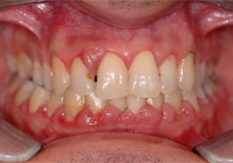 Figure 2: After 2 months of hormone therapy, without any periodontal treatment, gingival remission was observed.
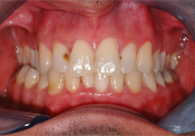 Figure 3: After 1 year, complete remission of the gingival hyperplasia was observed.
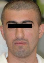 Figure 4: After a 1-year course of hormone treatment, the patient's weight returned to normal.
Conclusion
Dentists should be aware of a potential association between clinically observable gingival hyperplasia and hypothyroidism. Patients with this combination of symptoms should be referred to an endocrine clinic for definitive diagnosis, to prevent the potential complications of uncontrolled hypothyroidism.
THE AUTHORS
|
Dr. Fisekcioglu is an assistant professor in the department of oral diagnosis and radiology, faculty of dentistry, Yeditepe University, Istanbul, Turkey. |
|
|
Dr. Dolekoglu is an assistant professor in the department of oral diagnosis and radiology, faculty of dentistry, Yeditepe University, Istanbul, Turkey. |
|
|
Dr. Ilguy is an associate professor in the department of oral diagnosis and radiology, faculty of dentistry, Yeditepe University, Istanbul, Turkey. |
Correspondence to: Dr. Erdogan Fisekcioglu, Yeditepe University, Faculty of dentistry, Department of oral diagnosis and radiology, Bagdat Caddesi No: 238 Goztepe 34728 Istanbul, Turkey. Email: erdogan.fisekcioglu@yeditepe.edu.tr
The authors have no declared financial interests.
This article has been peer reviewed.
References
- Dongari-Baqtzoglou A; Research, Science and Therapy Committee, American Academy of Periodontology. Drug-associated gingival enlargement. J Periodontol. 2004;75(10):1424-31.
- Kinane DF, Marshall GJ. Periodontal manifestations of systemic disease. Aust Dent J. 2001;46(1):2-12.
- Persson RE, Hollender LG, MacEntee MI, Wyatt CC, Kiyak HA, Persson GR. Assessment of periodontal conditions and systemic disease in older subjects. J Clin Periodontol 2003;30(3): 207-213.
- Laine MA. Effect of pregnancy on periodontal and dental health. Acta Odontol Scand. 2002;60(5):257-64.
- Teng YT, Taylor GW, Scannapieco F, Kinane DF, Curtis M, Beck JD, et al. Periodontal health and systemic disorders. J Can Dent Assoc. 2002;68(3):188-92.
- Scardina GA, Messina P. Modifications of interdental papilla microcirculation: a possible cause of periodontal disease in Hashimoto's thyroiditis? Ann Anat. 2008;190(3):258-63. Epub 2008 Mar 4.
- Danese MD, Powe NR, Sawin CT, Ladenson PW. Screening for mild thyroid failure at the periodic health examination: a decision and cost-effectiveness analysis. JAMA. 1996;276(4):285-92
- Chaikin BS. Report of a case of fibromatosis of the gingivae associated with a hypothyroidism. Periodontics. 1965;3(6):306-9.