ABSTRACT
A simple appliance to replace an 8-year-old’s central incisor, which was decoronated after a trauma, is described. A natural tooth cantilevered pontic bonded to the adjacent central incisor may be an immediate solution. This appliance may be used when clinical conditions do not allow for the use of a conservative removable or fixed partial denture.
Introduction
When a child loses a maxillary permanent central incisor as a result of a traumatic injury—causing cervical fracture or avulsion without the possibility of reimplantation—or decoronation, a number of options exist to bridge the space created, depending on the condition of the surrounding dentition and the character of the patient. They include both removable and fixed appliances.1
The most common removable appliance is an acrylic plate holding a tooth to replace the missing one,2,3 attached to the maxillary first molars by 2 Adam’s clasps. Another option is a “flipper,” where the replacing tooth is held by 2 clasps on the adjacent abutment teeth.
A fixed appliance may be a partial denture in the form of a metal wire welded on the palatal side of 2 bands cemented to the maxillary molars, with the replacing tooth attached to the wire. Other fixed options include bonding a pontic to the adjacent teeth4,5 and a fibre-reinforced bridge.6
In any of these options, the replacing tooth may be the natural crown or an acrylic or composite-resin prosthesis.
When circumstances do not allow the use of these methods—e.g., if the patient refuses a removable appliance or the adjacent teeth cannot support a flipper or bonded appliance because they are not fully erupted or they have been severely traumatized—the dentist is challenged to provide a solution.
We describe a simple appliance to replace a maxillary central incisor in an extremely active and restless 8-year-old boy whose maxillary right central incisor was decoronated after a trauma.
Case Report
An 8-year-old boy presented after his maxillary permanent right central incisor (tooth 11) had been decoronated by a dentist (Fig. 1). Twelve months earlier, the tooth had been avulsed and reimplanted 1 hour later. The tooth was filled with calcium hydroxide, and signs of ankylosis had appeared. The day before the current visit, the same tooth was fractured horizontally at the cementoenamel junction, below the gingiva. Following this second trauma, decoronation was carried out: the pulp canal was widened, instrumentation over the root apex was performed, the fracture edges were rounded and the root was covered with gingival tissue and properly sutured. The purpose of the decoronation was to preserve the bone in the alveolus for future implant.7
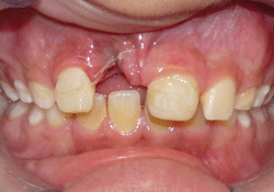 Figure 1: Frontal view immediately after decoronation of tooth 11.
The boy had an Angle Class II malocclusion, with a 4-mm overjet and 5-mm overbite, and was scheduled for orthodontic treatment after all permanent teeth were erupted. The lateral incisors were partly erupted. Thus, replacement of the decoronated tooth would be temporary, as the future treatment plan includes an implant through the resorbed root remaining in the alveolus.
As it was essential to maintain the space for the future orthodontic treatment and to do this immediately, options for replacing tooth 11 were discussed with the patient and his family. The patient refused a removable appliance. He desperately wanted a fixed appliance using his own tooth, but not the metal wire fixed partial denture described above. The option of bonding a pontic to both adjacent teeth (21 and 12) was rejected, as the depth of the overbite made a bilateral attachment difficult to achieve. In addition, the lateral incisor had not fully erupted. Thus, the pontic had to be bonded to tooth 21 only, creating a cantilevered pontic.8
The Appliance
During decoronation, the natural crown was saved to serve as the pontic. The crown was shortened to fit the incisogingival dimension without touching the gingiva to prevent irritation to the gingiva and to allow easy cleaning.7 The pulp chamber was removed and replaced with composite resin (Charisma, Heraeus Kulzer Gmbh, Hanau, Germany).
A 0.0175-inch (0.4445-mm) twist-flex wire (3M Unitek, Monrovia, CA), which is widely used for splinting after orthodontic treatment, was bent to form a 9-mm loop and to attach the pontic to the adjacent tooth (Fig. 2).9 The aim was to cover more than half the width of the pontic and the adjacent abutment incisor; the bend in the middle was made to fit the maxillary arch. To keep the pontic from touching the mandibular incisors after bonding, 0.3 mm of the enamel was removed from the palatal aspect of the decoronated crown.10,11 The loop was then bonded to the pontic using composite resin (Charisma) (Fig. 3). The pontic was held in place, adjusted to the arch and the free ends of the loop were bonded to the adjacent central incisor, tooth 21, with composite resin (Fig. 4).
 Figure 2: A twist-flex 0.0175-inch (0.4445-mm) wire bent into a 9-mm loop to attach the natural-tooth pontic to the adjacent central incisor.
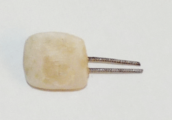 Figure 3: Buccal view of the loop bonded to the pontic with composite resin.
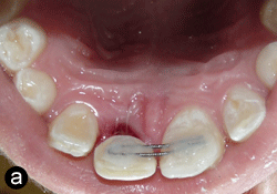
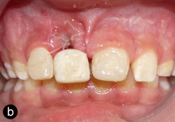
Figure 4: Palatal (a) and buccal (b) views of the pontic bonded to the adjacent central incisor with composite resin.
The patient was satisfied with the result (Fig. 5). However, the complexity of the solution was described to him, and he was warned to be careful when using the anterior teeth due to minor mobility of the pontic and of the need to maintain excellent oral hygiene.
The patient was seen at follow-up appointments after 1 week, then at 3-month intervals for a year. At the 6-month appointment, the decoronated area appeared normal on a radiograph, as did the lamina dura of tooth 21 (Fig. 6). Proper oral hygiene instructions were given at every follow-up appointment. During this whole period, the patient reported no eating problems, normal functioning and full satisfaction with the solution. Continuous eruption of tooth 12 was observed, along with some migration of this tooth into the space over the decoronated tooth (Fig. 7). After consultation with the patient, his family and the orthodontist, it was decided not to perform a second clinical procedure at this time, but postpone it until overall orthodontic treatment could be provided.
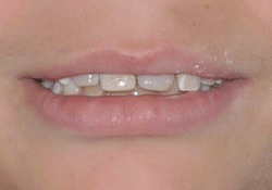 Figure 5: The resulting satisfactory esthetic appearance.
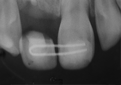 Figure 6: At the 6-month appointment, radiographic evaluation revealed a normal decoronated area and lamina dura of tooth 21
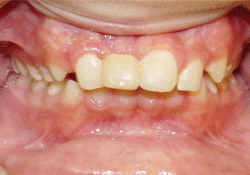 Figure 7: At 1-year follow-up, continuous eruption of tooth 12 had resulted in some migration into the space over the decoronated tooth.
Discussion
A cantilevered pontic attached to tooth 21 by a twist-flex wire loop was an immediate solution after decoronation as an interim measure until full orthodontic treatment could begin and reevaluation could take place. This appliance provided a satisfactory esthetic appearance after only a short chair time. The twist-flex wire allowed for some flexibility of the pontic, without creating loading forces on the adjacent central incisor, but also provided stability of the pontic. A narrow space was kept between the pontic and the gingiva to allow easy cleaning and continuous vertical bone growth at the site of decoronation.
This conservative approach, using a resin-bonded, twist-flex free cantilevered pontic, requires that the abutment tooth be intact or minimally restored, with substantial enamel present for bonding.12 These conditions existed in our case.
The successful use of resin-bonded prostheses requires establishment of retention.13 The extent of the overbite and the fact that the adjacent lateral incisor had not fully erupted prevented the use of the whole palatal surface of the tooth for bonding, but an effort was made to bond to the maximum palatal surface for maximum retention.
The natural-tooth pontic allowed good oral hygiene and reduced the risk of caries compared with fixed cemented appliances. It also provided a psychological benefit (it was the patient’s own tooth) and a fair esthetic match. However, it must be kept in mind that despite warnings to be careful when using the anterior teeth, an active 8-year-old boy has little control over biting movements of these teeth.
Regarding the suspected migration of tooth 12 into the space over the decoronated tooth, a second step to preserve the space was considered; however, the patient and parents preferred that this be carried out at the time of the full orthodontic treatment which was scheduled after all permanent teeth had erupted.
Removable partial dentures have the disadvantage of being dependent on patient compliance, and they may be lost or broken. In our case, the patient’s temperament prevented the use of a removable appliance.
Although not superior to a fixed partial denture in the form of a palatal wire welded on the palatal side of 2 bands cemented to the maxillary first molars, with the replacement tooth attached to the wire, the suggested appliance proved efficient and may expand the dentist’s arsenal of treatment options in selected cases.
THE AUTHORS
|
Dr. Peretz is an associate professor and head of the department of pediatric dentistry, Maurice and Gabriela Goldschleger School of Dental Medicine, faculty of medicine, Tel Aviv University, Tel Aviv, Israel. |
|
|
Dr. Nuni is a clinical instructor in the department of endodontics, Hebrew University-Hadassah faculty of dental medicine, Jerusalem, Israel. |
Correspondence to: Dr. Benjamin Peretz, Department of pediatric dentistry, Maurice and Gabriela Goldschleger School of Dental Medicine, Faculty of medicine, Tel Aviv University, Tel Aviv, Israel. Email: bperetz@post.tau.ac.il
The authors have no declared financial interests.
This article has been peer reviewed.
References
- Goodacre CJ, Cronin RJ Jr, Brown DT. Prosthodontic treatment of the adolescent patient. In: McDonald RE, Avery DR, Dean JA, editors. Dentistry for the child and adolescent. 8th ed. St. Louis: CV Mosby; 2004. p. 509-15.
- Finn SB. Clinical pedodontics. 4th ed. Philadelphia: WB Saunders; 1973. p. 259-60.
- Dyson JE. Prosthodontics for children. In: Wei SHY, editor. Pediatric dentistry — total patient care. Philadelphia: Lea & Febiger; 1988. p. 263.
- Graber TM, Swain BF. Orthodontics: current principles and techniques. 3rd ed. St. Louis: CV Mosby; 1985. p. 224-7.
- Sharma U, Garg AK, Gauba K. An interim, fixed prosthesis using natural tooth crown as a pontic. Contemp Clin Dent. 2010;1(2):130-2.
- Chafaie A, Portier R. Anterior fiber-reinforced composite resin bridge: a case report. Pediatr Dent. 2004;26(6):530-4.
- Malmgren B. Decoronation: why, how and when? J Calif Dent Assoc. 2000;28(11):846-54.
- Hussey DL. Pagni C, Linded GJ. Performance of 400 adhesive bridges fitted in a restorative dentistry department. J Dent. 1991;19(4):221-5.
- Zachrisson BU, Buyukyilmaz T. Bonding in orthodontics. In: Graber TM, Vanarsdall RL Jr, Vig KW, editors. Orthodontics: current principles and techniques. 4th ed. St Louis: Elsevier Mosby; 2005. p. 621-44.
- Al-Diwani H, Hyde TP, Gregory P, Brunton P. Providing support for the pontic of natural tooth adhesive bridges: a clinical report. Eur J Prosthodont Restor Dent. 2010;18(3):128-31.
- Botelho M. Resin-bonded prostheses: the current state of development. Quintessence Int. 1999;30(8):525-34.
- Pegoraro LF, Barrack G. A comparison of bond strengths of adhesive cast restorations using different designs, bonding agents, and luting resins. J Prosth Dent. 1987;57(2):133-8.
- Barrack G, Bretz WA. A long-term prospective study of the etched-cast restorations. Int J Prosthodont. 1993;6(5):428-34.