Successful orthodontic treatment depends on the orthodontist’s ability to control tooth movement and relies on a stable interface between wire and bracket.1 The adhesive system that bonds the bracket to enamel must be strong enough to resist all masticatory and orthodontic forces and remain adhered to the tooth and bracket throughout the course of treatment. In 1975, a shear bond strength (SBS) of 5.9–7.8 MPa for orthodontic brackets was first shown to be sufficient to withstand such forces.2 Although empirically established, this is still the clinically accepted SBS range for orthodontic brackets.3-5 Although it is critical that the adhesive withstand such forces during treatment, on completion of treatment, debonding of brackets must occur with minimal patient discomfort and enamel damage.6
Currently in orthodontics, total-etch, multi-step adhesive systems (TEMSAS) are most commonly used to bond brackets to enamel. These systems provide adequate bond strength3 to withstand masticatory forces. However, bonding as well as debonding appointments are time consuming for both the orthodontist and the patient, and debonding can cause enamel damage. The demand by dental professionals for adhesives with reduced technique sensitivity, shorter clinical application time7,8 and lower incidence of post-operative sensitivity has led to the development of self-etching adhesive systems.9,10 Among these are universal self-etch 1-step adhesive systems (USE1SASs) that combine the 3 steps required for adhesion into a 1-step application. These systems can be used in self-etch mode, selective enamel-etch mode or total-etch mode for restorative procedures. When used in a self-etch mode, USE1SASs may significantly simplify the bonding process by reducing the number of bonding steps and eliminating the need for total acid etching.11 In turn, this would decrease the risk of contamination, reduce the bonding procedure time11 and, potentially, reduce the risks of damaging enamel during debonding.
USE1SASs have been tested extensively on dentin and enamel surfaces. Although the SBSs reported in many of these studies are within the recommended 5.9–7.8 MPa2 for orthodontics, most were measured on cut enamel.5,12 Only limited studies exist on the performance of self-etch adhesives on uncut enamel13 or in an orthodontic setting.8
The objectives of this study were to investigate the SBS and debonded enamel surface characteristics of 3 USE1SASs, compared with a TEMSAS, used in bonding orthodontic brackets to uncut enamel at 2 time points. The tested null hypotheses were: there are no significant differences in SBSs among tested bonding agents; there are no significant differences in SBSs after a 6-month aging period; there are no significant differences among tested bonding agents in the amount of remaining resin on teeth after bracket debonding.
Material and Methods
Experimental Design
We studied 4 bonding agents at baseline and 6 months, using 8 groups of 20 teeth. The quantitative response variables were enamel SBS, adhesive remnant index (ARI) score and evaluation of remaining resin (%RR). Scanning electron microscopy (SEM) was used for qualitative analysis of debonded surfaces.
Materials Used
The composition of the materials used in this study is shown in Table 1.
| Adhesive | Composition |
|---|---|
| Note: bis-GMA = bisphenol A-glycidyl methacrylate, HEMA = hydroxy ethyl methacrylate, MDP = 10-methacryloyloxydecyl dihydrogen phosphate. |
|
| Scotchbond Universal Adhesive (pH = 2.7) | MDP; dimethacrylate resins; HEMA; Vitrebond copolymer filler; ethanol; water; initiators; silane |
| All-Bond Universal (pH = 3.2) | MDP; bis-GMA; HEMA; ethanol; water; initiators |
| Clearfil Universal Bond (pH = 2.3) |
bis-GMA; HEMA; MDP; gydrophilic aliphatic dimethacrylate; colloidal silica; dl-camphorquinone; silane coupling agent; zirconium oxide; accelerators; initiators; water; ethanol |
| Adper Scotchbond Multi-Purpose Adhesive | Etchant: 35% H3PO4; primer: HEMA, polyalkenoic acid polymer, water; adhesive resin: bis-GMA, HEMA, initiators |
| Transbond XT Light Cure Adhesive | Silane treated quartz; bisphenol A diglycidyl ether dimethacrylate; bisphenol A bis(2-hydroxyethyl ether) dimethacrylate; silane treated silica; diphenyliodonium hexafluorophosphate |
Definition of Groups and Sample Preparation
A total of 160 extracted, caries-free human premolars were used in the study. Tooth inclusion criteria included absence of endodontic treatment, carious lesions, restorations and enamel defects, such as enamel hypoplasia, enamel hypomineralization or visible cracks. The selected teeth were disinfected in 0.5% chloramine-T solution for 1 week, stored in distilled water at 37°C and used within 6 months of extraction. Teeth were randomly divided into 4 groups, based on the adhesive to be used: Scotchbond Universal (SU; 3M ESPE, St. Paul, MN, USA ); All-Bond Universal (BU; Bisco Dental Products, Schaumburg, IL, USA); Clearfil Universal Bond (CU; Kuraray Dental, New York, NY, USA); and the control (C), which was Adper Scotchbond Multi-Purpose (3M ESPE, St. Paul, MN, USA).
All teeth were initially cleaned and pumiced using a rubber cup with fluoride-free paste for 10 s, thoroughly washed with water and air dried. Bonding procedures were performed according to the manufacturers’ recommendations. Light-polymerization was performed using an Ortholux Luminous Curing Light (3M Unitek, Monrovia, CA, USA) at 1600 mW/cm2 irradiance intensity.
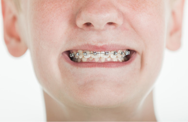
Stainless steel premolar brackets (Mini Master Series, 0.56 mm slot, surface area 10.29 mm2; American Orthodontics, Sheboygan, WI, USA) with the best surface contact and fit on the buccal surface of the teeth were chosen. Retention pads provided dual mechanical retention by layering 80-gauge mesh over an etched foil base. To bond the brackets, Transbond XT Light Cure Adhesive (3M Unitek) was compressed with a plastic instrument into the mesh of the brackets, which were then placed on the buccal surface of the tooth, with their slots parallel to the incisal edge. After pressing the brackets onto the buccal surfaces of the teeth with a carver instrument and gentle removal of any excess adhesive, the curing light was held stationary at a distance of 1–2 mm from the bracket for 12 s, with the light beam directed for 6 s each at the mesial and distal aspects of the bracket.14
The 40 teeth from each adhesive group were then divided into 2 groups (n = 20) and stored for 24 h (baseline) or
6 months in 37°C distilled water.
Shear Bond Strength
Before testing SBS, each tooth was placed in a circular mounting jig (made of SR Ivolen’s polymethyl methacrylate base; Ivoclar Vivadent, Schaan, Liechtenstein) for consistent tooth alignment such that an occluso-gingival load could be applied by a chisel to produce a shear force at the bracket–tooth interface. To ensure that the chisel blade attached to the Universal Testing Machine (model 4301; Instron, Norwood, Mass., USA) contacted each bracket from the incisal aspect as close to the bonding interface as possible, the facial surface of the tooth was mounted parallel to the chisel. Each bracket was then debonded at a crosshead speed of 1 mm/minute. The maximum force required to debond a bracket was recorded and mean SBS (in MPa) was calculated for each group.
Adhesive Remnant Index and Quantitative Evaluation of Remaining Resin
After bracket debonding, the enamel surface of each tooth was examined under a stereomicroscope at 10× magnification. Tested surfaces were classified according to the ARI scores described by Artun and Bergland.15 The ARI is a scaled score based on the amount of adhesive left on the enamel surface: 0 = none, 1 and 2 = less than half and greater than half, respectively, and 3 = all (Fig. 1). The frequency of each score was recorded and results were expressed as the percentage of each score for each adhesive and time.

Figure 1: Stereomicroscopic images (10×) of enamel surfaces. A: All-Bond Universal (BU) group at 6 months, adhesive remnant index (ARI) score = 0. B: Clearfil Universal Bond (CU) group at baseline, ARI score = 1. C: control (Adper Scotchbond Multi-Purpose Adhesive) group at 6 months, ARI score = 2. D: control group at 6 months, ARI score = 3.
In addition, the amount of adhesive remaining on teeth was analyzed by quantitative measurements from enlarged images of the tooth surfaces. Individual photographs of debonded surfaces (alongside a ruler) were taken with a Spot Insight Color 3.2.0 Camera (Diagnostic Instruments Inc., Sterling Heights, MI, USA) and the surface area of the bracket and remaining resin for each tooth was measured using ImageJ Software (National Institutes of Health, Bethesda, Md., USA). The residual resin for each tooth was expressed as a percentage of the remaining resin within the bracket perimeter (%RR).
Scanning Electron Microscopy
Samples representing each ARI score were qualitatively analyzed with SEM (Leica EM ACE200, Leica Microsystems, Wetzlar, Germany) at 500× and 1000× magnification.
Statistical Analysis
Two-way analysis of variance (ANOVA) and the Tukey post hoc test were used to compare mean SBS and %RR among the groups (α = 0.05).
Ethics approval
Ethics approval for this study was obtained from the University of Toronto research ethics board (protocol #31823).
Results
Bond Strength
For all tests, the assumption of normal distribution of errors was checked and satisfied by the Shapiro-Wilk test. The evaluated factors — group (p = 0.000), time (p = 0.001) and group × time (p = 0.009) — had a significant effect on SBS. The BU and C groups had the lowest and highest SBS values, respectively (p < 0.05), at both baseline and 6 months (Table 2). At both times, SU and CU had similar results and did not differ significantly from each other (p > 0.05). Significantly lower mean SBS was observed at 6 months compared with baseline for C, but there was no significant difference over time for the USE1SASs. SBS of USE1SASs ranged from 1.9 to 4.1 MPa at baseline and 0.55 to 4.6 MPa at 6 months; these SBS values are not considered appropriate for orthodontic treatment.
| Time | Shear bond strength, mean MPa ± SD | |||
|---|---|---|---|---|
| SU | BU | CU | C | |
| *SU = Scotchbond Universal Adhesive, BU = All-Bond Universal, CU = Clearfil Universal Bond, C = control (Adper™ Scotchbond Multi-Purpose Adhesive).Note: Different uppercase letters within a row indicate significant differences among means. Different lowercase letters within a column indicate significant differences among means. |
||||
| Baseline | 3.8 ± 2.0ABa | 1.9 ± 1.0Ab | 4.1 ± 1.4Bc | 8.4 ± 3.4Cd |
| 6 months | 2.9 ± 1.0Aa | 0.55 ± 0.35Bb | 4.6 ± 2.3ACc | 6.0 ± 2.0Ce |
Remaining Resin and Surface Characteristics of Enamel after Debonding
All 3 USE1SASs showed ARI scores of 0 or 1 at both times, indicating that there was no resin or less than half of the resin left on the enamel. A larger number of teeth in the control group showed remaining resin, especially at 6 months. At baseline, the ARI scores for the 3 USE1SASs were similar. However, for group C, 84% of teeth had a score of 1 and 16% had a score of 2 (Table 3). Although the most predominant mode of failure for all the adhesive groups at baseline and 6 months (Table 4) was a combination of adhesive and cohesive failures, there was a trend toward less resin remaining on the tooth surface in the USE1SAS groups compared with the control at 6 months.
The trend in ARI scoring was confirmed by the more precise quantification of remaining adhesive on enamel surfaces after debonding (Table 5). All evaluated factors — group, time and group × time (p = 0.000) — had a significant effect on %RR. The C group had a significantly higher mean percentage of remaining resin on the tooth surfaces compared with the 3 USE1SASs at both times (p < 0.05), with no significant differences among the self-etching adhesives. Comparing differences at baseline and 6 months, C and BU groups showed a significant difference (p < 0.001).
| Adhesive system* | ARI score, %† | |||
|---|---|---|---|---|
| 0 | 1 | 2 | 3 | |
| *SU = Scotchbond Universal Adhesive, BU = All-Bond Universal, CU = Clearfil Universal Bond, C = control (Adper™ Scotchbond Multi-Purpose Adhesive).Note: Different uppercase letters within a row indicate significant differences among means. Different lowercase letters within a column indicate significant differences among means. |
||||
| SU | 0 | 100 | 0 | 0 |
| BU | 0 | 100 | 0 | 0 |
| CU | 0 | 100 | 0 | 0 |
| C | 0 | 84 | 16 | 0 |
| ARI score, %† | ||||
|---|---|---|---|---|
| 0 | 1 | 2 | 3 | |
| *SU = Scotchbond Universal Adhesive, BU = All-Bond Universal, CU = Clearfil Universal Bond, C = control (Adper Scotchbond Multi-Purpose Adhesive). †Amount of adhesive left on tooth: 0 = none, 1 = less than half, 2 = more than half, 3 = all. | ||||
| SU | 6 | 94 | 0 | 0 |
| BU | 17 | 83 | 0 | 0 |
| CU | 0 | 100 | 0 | 0 |
| C | 0 | 6 | 76 | 18 |
| Time | Remaining adhesive, mean % ± SD | |||
| SU | BU | CU | C | |
| *SU = Scotchbond Universal Adhesive, BU = All-Bond Universal, CU = Clearfil Universal Bond, C = control (Adper Scotchbond Multi-Purpose Adhesive). Note: Different uppercase letters within a row indicate significant differences among means. Different lowercase letters within a column indicate significant differences among means. | ||||
| Baseline | 7.0 ± 4.9Aa | 7.2 ± 4.6Ab | 11.4 ± 6.7Ad | 30.4 ± 20.7Be |
| 6 months | 6.4 ± 5.3Ya | 1.7 ± 1.6Yc | 9.4 ± 6.6Yd | 77.4 ± 18.1Zf |
SEM analyses of the debonded enamel surfaces showed that, in general, the enamel surface of teeth in the USE1SAS groups appeared smooth and less porous with minimal remaining resin on the enamel surfaces (Figs. 2 and 3). In contrast, for the C group, superficial microporosities are clearly visible on total-etched debonded surfaces.
Based on our results, all 3 null hypotheses were rejected.

Figure 2: Scanning electron microscope images (1000×) of debonded surfaces at baseline. Note: E = enamel, R = resin. A: Scotchbond Universal Adhesive (SU) group. B: All-Bond Universal (BU) group. C: Clearfil Universal Bond (CU) group. D: control (Adper Scotchbond Multi-Purpose Adhesive) group.

Figure 3: Scanning electron microscope images (500×) of debonded surfaces at 6 months. Note: AD = adhesive, E = enamel, R = resin. A: Scotchbond Universal Adhesive (SU) group. B: All-Bond Universal (BU) group. C: Clearfil Universal Bond (CU) group. D: control (Adper Scotchbond Multi-Purpose Adhesive) group.
Discussion
Mild or ultra-mild USE1SASs (pH 2.3–3.2), such as the ones used in this study, rely on a 2-fold bonding mechanism: a micromechanical bond and a chemical bond.7 Their monomers usually contain a carboxylic or phosphoric acid group, which etches the tooth, creating surface porosity to produce mechanical retention.7 The chemical bond is related to the presence of a specific functional monomer, 10-methacryloyloxydecyl dihydrogen phosphate (10-MDP), which combines enamel demineralization with the ability to bond ionically with the calcium ions of hydroxyapatite.16,17 Uncut enamel is a hypermineralized and aprismatic substrate by nature.13 On such a substrate, this study showed that the more aggressive demineralizing effect created by the TEMSAS (H3PO4, pH < 1) superseded the dual bonding mechanism of the 3 USE1SASs, leading to significantly higher SBS.
The other factor that influences SBS is the presence of functional monomer impurities that can affect the durability of the enamel bond.18,19 Yoshihara and colleagues18 confirmed that both the purity and presence of 10-MDP dimers in adhesives influence the etching efficacy of hydroxyapatite and bond strength.Therefore, it is possible that the lower SBS of BU compared with SU and CU may be a result of impurities in the 10-MDP functional monomer. These impurities and dimers may undergo hydrolytic degradation more rapidly, thus accounting for the decrease in SBS with time. Although no significant individual decrease in SBS in the USE1SAS groups occurred after 6 months, our results indicate that the factor time did significantly reduce overall SBS (p = 0.001). These results, especially in the CU group, are in agreement with McLean et al.12 and Atash Biz Yeganeh et al.20
Studies have reported a relation between bond strength and failure mode, as greater bond strengths correlate with more mixed fractures.21 This relation was apparent when we looked at bond strength and the amount of resin remaining after debonding in the C group compared with the USE1SAS groups at both times. These results are in agreement with Sharma et al.,5 who showed that a total-etch system had a greater ARI score than self-etch systems. In addition, Schnebel et al.22 showed that total-etch adhesives fail mainly at the bracket–adhesive interface, thus leaving more residual resin on the enamel surface. On the other hand, self-etch adhesives result in more failures at the enamel–adhesive interface, leaving less resin on the tooth surface. As bracket failure occurs at the weakest interface, this also indicated a weaker bond to the enamel surface, resulting in lower SBS.22
Our study shows a clear relation between SBS, ARI and %RR. Over time, the overall ARI score, %RR and SBS decreased for BU. A lower ARI score after 6 months signified less resin remaining on the tooth and a weaker bond between the resin and the enamel, which correlated with its decrease in SBS. Although the overall ARI score and SBS also decreased for the SU group, there was no difference in %RR. An ARI score of 1 remained constant for CU and there was no difference in %RR, which reflected a greater stability of SBS. However, for the C group, although the overall ARI score and mean %RR increased over the 6-month period, there was a decrease in SBS. A study by Burrow et al.,23 which assessed the 7-year dentin bond strength of a total-etch and a self-etch system, demonstrated similar results. In that study, although the SBS of both systems decreased over time, the mode of failure for the self-etch system did not change with time, whereas the total-etch system had an increase in cohesive failures in dentin.23 This signifies that, in the present study, the bond between the resin and tooth, for some samples, may have increased over time and the weakest point was between the bracket and adhesive system. In general, the %RR results of this study are in agreement with the ARI scores, thus validating the ARI scoring index. In a comparative study of qualitative and quantitative methods for the assessment of adhesive remnant after bracket debonding, Cehreli et al.24 also concluded that qualitative visual scoring using the ARI is capable of generating results that are consistent with those assessed by quantitative image analysis techniques.
Under SEM, the amount of residual resin on selected enamel surfaces was greater than the calculated percentage of remaining resin; however, there was no change in ARI score for the selected teeth. As previously mentioned, enamel surface conditioning by the USE1SASs was less effective than TEMSAS conditioning, possibly resulting in decreased micromechanical retention. Figures 1 and 2 show that enamel surfaces in the USE1SAS groups were smooth after debonding and less porous than surfaces in the TEMSAS group. Although of relevance to orthodontics, not many studies have evaluated the strength of bonding agents to uncut enamel. In 2003, Perdigao and Geraldeli25 evaluated immediate bond strength of 1-step and 2-step self-etch adhesives, compared with a 2-step total-etch adhesive, on uncut enamel. Microtensile bond strength for 1-step and 2-step self-etch adhesives was 0.08–11.8 MPa and 11.3–16.7 MPa, respectively, whereas the 2-step total-etch adhesive had the highest bond strength at 31.5 MPa. The very low mean bond strength value obtained for the One-Up Bond F system (Tokuyama Dental Corp., Taitou-ku Tokyo, Japan), i.e., 0.08 MPa, was justified by its “relatively high pH of 2.57.” These results were supported by field emission SEM analyses, which showed virtually no interprismatic penetration of the adhesive as well as formation of gaps across the entire interface. In another study, Patil et al.13 found that the immediate bond strength of 2-step total-etch was 4.94 MPa compared with 3.62 MPa for 1-step self-etch bonding agent to uncut enamel. The range for SBS for USE1SASs in the present study (immediate 1.9–4.1 MPa, aged 0.55–4.6 MPa) falls within and is consistent with previous literature. Differences in absolute mean values may be attributed to variations in the chemistry of bonding systems, testing methods and bonding substrate, such as age of teeth and fluoridation.
This study showed that USE1SASs not only had significantly lower SBS to uncut enamel than the TEMSAS, but also that SBS values were below minimal strength appropriate for orthodontic applications in self-etch mode.
THE AUTHORS
 |
Dr. Cerone is former orthodontics resident, department of clinical sciences (orthodontics), faculty of dentistry, University of Toronto, Toronto, Ontario. |
 |
Dr. El-Badrawy is associate professor, department of clinical sciences (restorative), faculty of dentistry, University of Toronto, Toronto, Ontario. |
 |
Dr. Gong is associate professor, department of clinical sciences (orthodontics), faculty of dentistry, University of Toronto, Toronto, Ontario. |
 |
Dr. Prakki is associate professor, department of clinical sciences (restorative), faculty of dentistry, University of Toronto, Toronto, Ontario. |
Correspondence to: Dr. Anuradha Prakki, Associate Professor, Faculty of Dentistry, University of Toronto, 124 Edward St, Room 537, Toronto, ON M5G 1G6. anuradha.prakki@dentistry.utoronto.ca
The authors have no declared financial interests.
This article has been peer reviewed.
References
- Costa AR, Vedovello-Filho M, Correr AB, Vedovello SA, Puppin-Rontani RM Ogliari FA, et al. Bonding orthodontics brackets to enamel using experimental composites with an iodonium salt. Eur J Orthod. 2014;36(3):297-302.
- Reynolds I. A review of direct orthodontic bonding. Br J Orthod. 1975;2(3):171-8.
- Bakhadher W, Halawany H, Talic N, Abraham N, Jacob V. Factors affecting the shear bond strength of orthodontic brackets - a review of in vitro studies. Acta Medica (Hradec Kralove) 2015;58(2):43-8.
- Finnema KJ, Ozcan M, Post WJ, Ren Y, Dijkstra PU. In-vitro orthodontic bond strength testing: a systematic review and meta-analysis. Am J Orthod Dentofacial Orthop. 2010;137(5):615-22.e3.
- Sharma S, Tandon P, Nagar A, Singh GP, Singh A, Chugh VK. A comparison of shear bond strength of orthodontic brackets bonded with four different orthodontic adhesives. J Orthod Sci. 2014;3(2):29-33.
- Patil P, Kaur S, Kaur M, Kaur M, Vinuta S, Kaur RK. Orthodontic cements and adhesives: a review. J Adv Med Dent Sci Res. 2014;2(3):35-8.
- Giannini M, Makishi P, Ayres AP, Vermelho P, Fronza B, Nikaido T, et al. Self-etch adhesive systems: a literature review. Braz Dent J. 2015;26(1):3-10.
- Shaik JA, Reddy RK, Bhagyalakshmi K, Shah MJ, Madhavi O, Ramesh SV. In vitro evaluation of shear bond strength of orthodontic brackets bonded with different adhesives. Contemp Clin Dent. 2018;9(2):289-92.
- Christensen GJ. Self-etching primers are here. J Am Dent Assoc. 2001;132(7):1041-3.
- Stape THS, Tjäderhane L, Abuna G, Sinhoreti MAC, Martins LRM, Tezvergil-Mutluay A. Optimization of the etch-and-rinse technique: new perspectives to improve resin–dentin bonding and hybrid layer integrity by reducing residual water using dimethyl sulfoxide pretreatments. Dent Mater. 2018;34(7):967-77.
- Nair M, Paul J, Kumar S, Chakravarthy Y, Krishna V, Shivaprasad. Comparative evaluation of the bonding efficacy of sixth and seventh generation bonding agents: an in-vitro study. J Conserv Dent. 2014;17(1):27-30.
- McLean DE, Meyers EJ, Guillory VL, Vandewalle KS. Enamel bond strength of new universal adhesive bonding agents. Oper Dent. 2015;40(4):410-7.
- Patil D, Singbal KP, Kamat S. Comparative evaluation of the enamel bond strength of ‘etch-and-rinse’ and ‘all-in-one’ bonding agents on cut and uncut enamel surfaces. J Conserv Dent. 2011;14(2):147-50.
- Swanson T, Dunn WJ, Childers DE, Taloumis LJ. Shear bond strength of orthodontic brackets bonded with light-emitting diode curing units at various polymerization times. Am J Orthod Dentofacial Orthop. 2004;125(3):337-41.
- Artun J, Bergland S. Clinical trials with crystal growth conditioning as an alternative to acid-etch enamel pretreatment. Am J Orthod. 1984;85(4):333-40.
- Cardoso MV, de Almeida Neves A, Mine A, Coutinho E, Van Landuyt K, De Munck J, et al. Current aspects on bonding effectiveness and stability in adhesive dentistry. Aust Dent J. 2011;56(Suppl 1):31-44.
- Van Meerbeek B, De Munck J, Yoshida Y, Inoue S, Vargas M, Vijay P, et al. Buonocore memorial lecture. Adhesion to enamel and dentin: current status and future challenges. Oper Dent. 2003;28(3):215-35.
- Yoshihara K, Nagaoka N, Okihara T, Kuroboshi M, Hayakawa S, Maruo Y, et al. Functional monomer impurity affects adhesive performance. Dent Mater. 2015;31(12):1493-501.
- Tsuchiya K, Takamizawa T, Barkmeier WW, Tsubota K, Tsujimoto A, Berry TP, et al. Effect of a functional monomer (MDP) on the enamel bond durability of single-step self-etch adhesives. Eur J Oral Sci. 2016;124(1):96-102.
- Atash Biz Yeganeh L, Seyed Tabai E, Mohammadi Basir M. Bonding durability of four adhesive systems. J Dent (Tehran). 2015;12(8):563-70.
- al-Salehi SK, Burke FJ. Methods used in dentin bonding tests: an analysis of 50 investigations on bond strength. Quintessence Int. 1997;28(11):717-23.
- Schnebel B, Mateer S, Maganzini AL, Freeman K. Clinical acceptability of two self-etch adhesive resins for the bonding of orthodontic brackets to enamel. J Orthod. 2012;39(4):256-61.
- Burrow MF, Harada N, Kitasako Y, Nikaido T, Tagami J. Seven-year dentin bond strengths of a total- and self-etch system. Eur J Oral Sci. 2005;113(3):265-70.
- Cehreli SB, Polat-Ozsoy O, Sar C, Cubukcu HE, Cehreli ZC. A comparative study of qualitative and quantitative methods for the assessment of adhesive remnant after bracket debonding. Eur J Orthod. 2012;34(2):188-92.
- Perdigão J, Geraldeli S. Bonding characteristics of self-etching adhesives to intact versus prepared enamel. J Esthet Restor Dent. 2003;15(1):32-42.